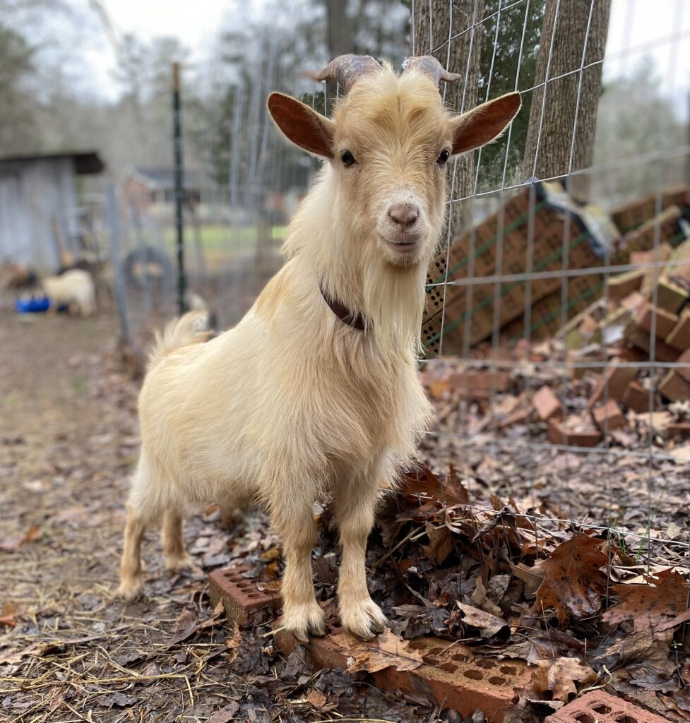
Urinary Calculi and Blockage in Male Goats

*Names and Images are changed for patient privacy*
Late in the afternoon a new owner called concerned that her 3 year old male castrated Nigerian dwarf
goat, “Prince”, was standing in his pen with “hunched” posture, and showing lethargy, dribbling urine, and decreased appetite for approximately the past 24 hours.
(These concerns noticed by the owner are buzz words for a goat in medical emergency who needs urgent evaluation!)
Owner also stated his diet consists of alfalfa hay and sweet feed.
On physical exam he was quiet, alert, responsive. Mucus membranes were pale pink and moist. CRT<2.
Heart and respiratory rates were within normal limits. 1 rumen contraction per minute. Abdomen was
tense and mildly painful to palpation. Urethral process (pizzle) was red, inflamed, necrotic.
(These are classic clinical signs of a urethra blocked with a stone preventing urination.)
(Prolonged blockages can cause the bladder to rupture which is always a surgical emergency. What
would be the best way to investigate if the bladder was ruptured?……….Ultrasound!)
Ultrasound showed a large, firm bladder with wall intact.
Goats have two places urethral stones commonly get stuck; sigmoid flexure and pizzle. At both of these
places the urethra is significantly more narrow. Obstructions can happen at one or both places
simultaneously with obstructions at multiple locations being most common. Unfortunately, obstructions
at the pizzle only have the best outcomes with medical management alone as obstructions higher up
can not be reached. This means medical management alone has much lower success rate compared to
surgical management.
At this time treatment options were discussed with the owner. If obstruction was only at pizzle, there
was a small percent chance of successfully removing obstruction with a pizzle amputation. There was a
much higher percent chance of multiple obstructions that would require surgical management at
university veterinary hospital. Pros and cons of both treatment options were discussed with owner.
(This owner opted to try medical management and if unsuccessful, would take to hospital)
Treatment: Administered sedation —> Amputated urethral process—> Inserted catheter into urethra in
which a fine stream of urine was produced —> Gently applied pressure to express bladder.
Gave injection of pain medication. Left the following medications: 7 days of pain and anti-inflammatory
tablets and 30 days ammonium chloride.
(Ammonium chloride is used to acidify the urine, which
decreases the ability to form stones. Long term continued use allows the body to neutralize the
ammonium chloride, so it should be dosed as needed on the recommendation of your veterinarian, not
continuously used.)
Urinary stones are most common in male castrated small ruminants fed high grain diets which can have
elevated levels of minerals such as calcium, phosphorus, and magnesium. (Remember this goat’s diet
consisted of sweet feed and alfalfa.) It’s important to keep calcium to phosphorus ratio in a diet less
then 2:1. These stones originally form in the bladder and then excreted in the urine through the urethra
where they can become “stuck” at the sigmoid flexure and/or urethral process. These areas are much
more narrow than the rest of the urethra.
Urethral stones causing blockages are EMERGENCIES! Common clinical signs to monitor for include
blood in urine, straining to urinate, decreased to absent urine production, abdominal pain, lethargy, decreased to no appetite, and swelling around prepuce. If you notice these clinical signs, call
your veterinarian!
Many thanks to Future Doctor Ridenour for this case summary!
